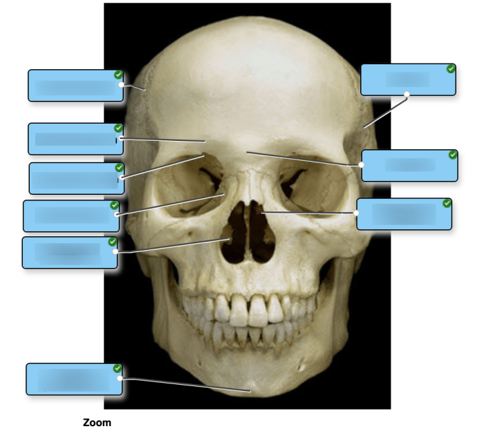
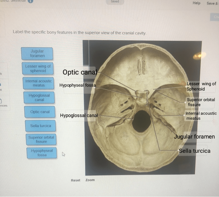
Label the specific bony features of the superior skull – Labeling the specific bony features of the superior skull is a fundamental aspect of understanding human anatomy. This intricate structure, composed of several bones, plays a vital role in protecting the brain, facilitating sensory functions, and providing structural support to the face.
Embark on a journey of discovery as we delve into the intricacies of the superior skull, unraveling its anatomical landmarks and their significance.
From the prominent frontal bone to the complex temporal bones, each component of the superior skull serves a unique purpose. We will explore the external and internal features of these bones, examining their sutures, foramina, and processes. Along the way, we will uncover the clinical significance and functions associated with each structure, providing a comprehensive understanding of this remarkable part of the human body.
Overview of the Superior Skull: Label The Specific Bony Features Of The Superior Skull

The superior skull, also known as the calvaria or cranium, forms the upper portion of the skull and protects the brain. It consists of eight bones that are tightly connected by sutures. These bones are the frontal, parietal, temporal, occipital, sphenoid, ethmoid, nasal, and lacrimal bones.
The study of the superior skull, known as craniology, has a long history, dating back to ancient times. It has played a crucial role in understanding human evolution, anatomy, and pathology.
Frontal Bone
The frontal bone is located at the anterior part of the superior skull and forms the forehead. It is bounded by the parietal bones posteriorly, the sphenoid bone inferiorly, and the nasal and lacrimal bones medially. Externally, the frontal bone presents the supraorbital margin, which forms the upper border of the orbits.
Internally, it contains the frontal sinuses, air-filled cavities that reduce the weight of the skull. The crista galli, a midline projection, articulates with the ethmoid bone and supports the olfactory bulb.
Parietal Bones
The parietal bones are paired bones that form the lateral and superior portions of the superior skull. They are bounded by the frontal bone anteriorly, the occipital bone posteriorly, and the temporal bones laterally. Externally, the parietal bones exhibit the sagittal suture, where they meet along the midline, and the lambdoid suture, where they meet the occipital bone.
Internally, they have the parietal foramina, which transmit blood vessels and nerves.
Temporal Bones
The temporal bones are complex and irregularly shaped bones located at the lateral and inferior parts of the superior skull. Externally, they present the mastoid process, which houses the mastoid air cells, the zygomatic process, which articulates with the zygomatic bone, and the external auditory meatus, the opening of the ear canal.
Internally, the temporal bones have the petrous part, which contains the inner ear, the middle ear, and the auditory ossicles.
Occipital Bone, Label the specific bony features of the superior skull
The occipital bone is located at the posterior part of the superior skull and forms the back of the head. It is bounded by the parietal bones anteriorly, the temporal bones laterally, and the sphenoid bone anteriorly. Externally, the occipital bone has the foramen magnum, through which the spinal cord passes, and the occipital condyles, which articulate with the atlas vertebra.
Internally, it exhibits the nuchal lines, which provide attachment for muscles.
Sphenoid Bone
The sphenoid bone is a complex and centrally located bone that forms the middle part of the base of the skull. It is bounded by the frontal bone anteriorly, the occipital bone posteriorly, the temporal bones laterally, and the ethmoid bone anteriorly.
Externally, the sphenoid bone has the sella turcica, which houses the pituitary gland, and the optic canals, which transmit the optic nerves. Internally, it contains the pterygoid processes, which provide attachment for muscles.
Ethmoid Bone
The ethmoid bone is a small and delicate bone located at the anterior part of the base of the skull. It is bounded by the frontal bone posteriorly, the sphenoid bone posteriorly, the nasal bones anteriorly, and the lacrimal bones laterally.
Externally, the ethmoid bone has the cribriform plate, through which the olfactory nerves pass, and the perpendicular plate, which forms part of the nasal septum. Internally, it contains the ethmoid sinuses, air-filled cavities that reduce the weight of the skull.
Nasal Bones
The nasal bones are paired bones that form the bridge of the nose. They are bounded by the frontal bone superiorly, the maxillae inferiorly, and the lacrimal bones laterally. Externally, the nasal bones exhibit the nasal bridge, which is the bony prominence of the nose, and the nasal septum, which divides the nasal cavity into two.
Lacrimal Bones
The lacrimal bones are small and thin bones located at the medial wall of the orbit. They are bounded by the frontal bone superiorly, the maxillae inferiorly, and the ethmoid bone posteriorly. Externally, the lacrimal bones have the lacrimal fossa, which houses the lacrimal sac, and the lacrimal groove, which transmits the nasolacrimal duct.
Zygomatic Bones
The zygomatic bones are paired bones that form the cheekbones. They are bounded by the frontal bone superiorly, the maxillae inferiorly, and the temporal bone posteriorly. Externally, the zygomatic bones have the zygomatic arch, which forms the lateral border of the orbit, and the temporal process, which articulates with the temporal bone.
Maxillae
The maxillae are paired bones that form the upper jaw. They are bounded by the frontal bone superiorly, the palatine bones posteriorly, and the zygomatic bones laterally. Externally, the maxillae have the alveolar process, which supports the teeth, and the maxillary sinus, an air-filled cavity that reduces the weight of the skull.
Internally, they contain the palatine process, which forms the hard palate.
Mandible
The mandible is a single bone that forms the lower jaw. It is bounded by the maxillae superiorly and the temporal bones laterally. Externally, the mandible has the body, which contains the alveolar process that supports the teeth, and the ramus, which articulates with the temporal bone.
Internally, it has the condylar process, which articulates with the temporal bone.
FAQ Overview
What is the largest bone in the superior skull?
The frontal bone is the largest bone in the superior skull.
Which bone forms the roof of the orbit?
The frontal bone forms the roof of the orbit.
What is the function of the temporal bone?
The temporal bone houses the organs of hearing and balance, and also contains the facial nerve.

